



Introduction
For hundreds of years, forensic doctors have followed one basic principle: to find out how a person died, the body must be examined through a traditional autopsy. This method has played an important role in solving crimes, identifying diseases, and delivering justice.
However, conventional autopsies have some important limitations. They involve surgical dissection of the body, which permanently changes it. They also expose doctors and forensic staff to infectious diseases and may conflict with the religious or cultural beliefs of the deceased’s family.
Today, advances in medical imaging and digital technology are offering a new approach. Virtual Autopsy (Virtopsy) allows forensic experts to examine the body without making large surgical cuts. Using advanced scanning techniques, it creates a detailed three-dimensional (3D) digital image of the body, helping investigators determine the cause of death with greater precision while preserving the dignity of the deceased.
What Is Virtopsy?
Virtopsy, also known as virtual autopsy, is a non-invasive method of examining a body after death. It was developed in the late 1990s at the Institute of Forensic Medicine in Bern, Switzerland.
Instead of using a scalpel, Virtopsy uses advanced imaging technologies to examine the body from both the outside and the inside.
Also Read: File Legal Notice in India: Complete Guide to Drafting, Procedure, Cost & Legal Rights
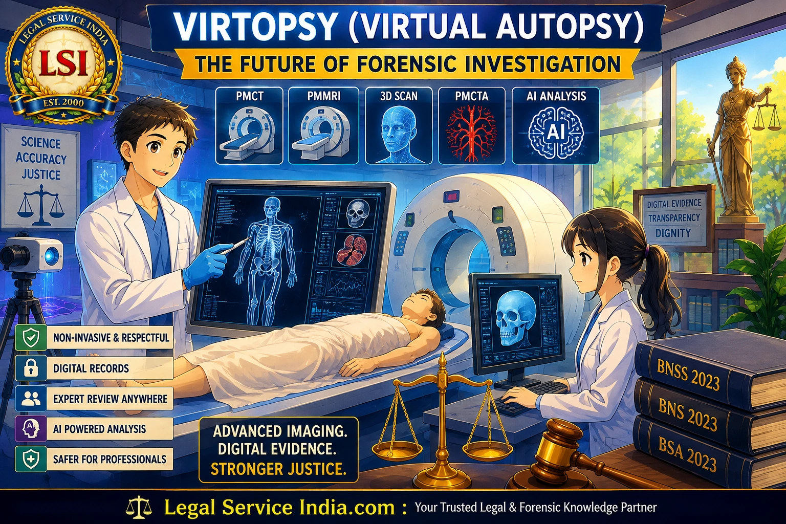
The main technologies used are
| Technology | Primary Purpose | Main Findings |
|---|---|---|
| Post-Mortem Computed Tomography (PMCT) | 3D imaging of bones and internal structures | Fractures, internal bleeding, bullet injuries, gas, foreign objects |
| Post-Mortem Magnetic Resonance Imaging (PMMRI) | Detailed soft tissue imaging | Brain injuries, organ damage, muscle injuries, heart diseases |
| 3D Surface Scanning | External body documentation | Bruises, wounds, bite marks, ligature marks |
| Post-Mortem CT Angiography (PMCTA) | Blood vessel examination using contrast dye | Hidden bleeding, damaged vessels, blocked arteries |
Main Technologies Used in Virtopsy
A. Post-Mortem Computed Tomography (PMCT)
PMCT uses CT scans to produce detailed 3D images of bones and internal structures. It helps detect:
- Bone fractures
- Bullet and knife injuries
- Foreign objects such as bullet fragments
- Internal bleeding
- Gas inside the body
- Major traumatic injuries
B. Post-Mortem Magnetic Resonance Imaging (PMMRI)
PMMRI provides clear images of soft tissues and internal organs. It is useful for examining:
- Brain injuries
- Heart diseases
- Organ damage
- Muscle injuries
- Injuries in children
C. 3D Surface Scanning
High-resolution cameras and optical scanners capture detailed images of the body’s surface. They record:
- Cuts and wounds
- Bruises
- Bite marks
- Ligature marks
- Entry and exit wounds
These images are combined with internal scans to provide a complete picture of the injuries.
D. Post-Mortem CT Angiography (PMCTA)
In some cases, a special contrast dye is injected into the blood vessels before the CT scan. This helps doctors identify:
- Hidden internal bleeding
- Damaged blood vessels
- Blocked arteries
- Heart-related conditions
Advantages of Virtopsy
A. Preserves the Dignity of the Deceased
Many religions and cultures believe that the body should remain intact after death. Since Virtopsy does not require major surgical dissection, it respects these beliefs while still allowing doctors to investigate the cause of death.
B. Provides Better Visualization
During a traditional autopsy, some natural relationships between organs and tissues may change once the body is opened.
Virtopsy records everything exactly as it appears inside the body before any examination begins. It can clearly show:
- Small bone fractures
- Internal injuries
- Bullet paths
- Organ damage
- Air and fluid inside the body
Because the scans are stored digitally, experts can examine them again whenever needed.
C. Creates Permanent Digital Records
A traditional autopsy cannot be repeated because the body changes after dissection.
Virtopsy creates permanent digital records in standard medical imaging format (DICOM). These records can be:
- Stored safely for many years
- Reviewed by other experts
- Shared with specialists anywhere in the world
- Re-examined if new evidence is found
- Used for teaching and research
This improves transparency and accuracy in forensic investigations.
D. Improves Safety for Forensic Professionals
Forensic doctors often work with bodies carrying infectious diseases such as
- Tuberculosis
- Hepatitis B and C
- HIV
- COVID-19
Because Virtopsy requires little or no direct contact with body tissues, it reduces the risk of infection and improves workplace safety.
E. Supports Artificial Intelligence (AI)
Modern Virtopsy systems are increasingly using Artificial Intelligence (AI) to assist forensic experts.
AI can help:
- Detect small fractures
- Measure internal bleeding
- Trace bullet paths
- Identify abnormal findings
- Speed up image analysis
Although final decisions are always made by forensic experts, AI makes the examination faster and more accurate.
Legal Importance in India
India’s new criminal laws—the Bharatiya Nagarik Suraksha Sanhita, 2023 (BNSS), Bharatiya Nyaya Sanhita, 2023 (BNS), and Bharatiya Sakshya Adhiniyam, 2023 (BSA)—place greater importance on scientific investigation and digital evidence.
Virtopsy fits well within this modern legal framework.
| Legal Aspect | Importance of Virtopsy |
|---|---|
| Scientific Investigation | Supports forensic-based investigations in serious criminal cases. |
| Digital Documentation | Creates permanent digital records without altering evidence. |
| Electronic Evidence | Digital images may qualify as electronic evidence under the BSA. |
| Courtroom Presentation | 3D images simplify the presentation of scientific evidence before courts. |
A. Scientific Investigation
The BNSS encourages greater involvement of forensic experts in serious criminal cases, helping investigations rely more on scientific evidence than on witness statements alone.
B. Digital Documentation
Virtopsy creates complete digital records of the body, which can be preserved without altering the original condition of the evidence.
C. Electronic Evidence
The Bharatiya Sakshya Adhiniyam recognizes electronic records as evidence in court, provided legal requirements regarding authenticity and integrity are met. Properly preserved Virtopsy images can therefore become valuable forensic evidence.
D. Easy Courtroom Presentation
Three-dimensional images are much easier for judges, lawyers, and juries to understand than traditional autopsy photographs.
They clearly show:
- Bullet trajectories
- Fractures
- Internal injuries
- Cause of trauma
This makes scientific evidence easier to explain in court.
Challenges of Virtopsy
Despite its many advantages, Virtopsy cannot completely replace the traditional autopsy.
Some investigations still require:
- Tissue examination under a microscope (histopathology)
- Toxicology tests
- DNA analysis
- Microbiological tests
- Chemical analysis for poisoning
Limitations of Virtopsy
In addition, Virtopsy requires expensive CT and MRI scanners, trained specialists, and advanced computer systems, making it difficult to implement everywhere.
| Challenge | Impact |
|---|---|
| Need for laboratory investigations | Some cases still require histopathology, toxicology, DNA, microbiology, and chemical analysis. |
| High infrastructure cost | Requires advanced CT and MRI scanners and specialized software. |
| Need for trained experts | Requires radiologists, forensic experts, and imaging specialists. |
| Limited availability | Not yet accessible in all hospitals and forensic laboratories. |
The Future: A Combination of Both Methods
Today, many countries follow a hybrid approach, combining Virtopsy with traditional autopsy.
A typical process is
- First, the body is examined using PMCT, PMMRI, and 3D surface scanning.
- If the scans clearly show the cause of death, no major surgical examination may be needed.
- If further investigation is required, doctors perform only limited and targeted tissue sampling or dissection based on the scan findings.
This approach improves accuracy while reducing unnecessary surgical procedures.
Benefits of the Hybrid Approach
- Improves diagnostic accuracy.
- Reduces unnecessary invasive procedures.
- Allows targeted tissue sampling.
- Combines digital imaging with laboratory confirmation.
- Enhances the reliability of forensic investigations.
Conclusion
Virtual autopsy is one of the most important advances in modern forensic science. It combines medical imaging, digital technology, and forensic medicine to make death investigations more accurate, safer, and more transparent.
It protects the dignity of the deceased, reduces health risks for forensic professionals, creates permanent digital evidence, and helps courts understand medical findings more clearly.
Although it cannot completely replace the traditional autopsy, Virtopsy is becoming an essential tool in modern forensic investigations. By combining advanced technology with conventional forensic methods, it is helping deliver more reliable scientific evidence and strengthening the administration of justice.
Key Takeaways
- Virtopsy offers a non-invasive alternative to many aspects of conventional autopsy.
- Certain investigations still require laboratory-based forensic examinations.
- A hybrid approach provides the best balance between imaging technology and traditional forensic methods.
- Digital evidence generated through Virtopsy improves transparency and courtroom presentation.
- Virtopsy is expected to play an increasingly important role in future forensic investigations.
Important Links
- Lawyers in India
- Copyright Registration in India
- Caveat Filing in Supreme Court of India
- Mutual Consent Divorce in Delhi NCR: WhatsApp 9650499965